Developing a Better Model
Feb 27, 2020 — Atlanta, GA

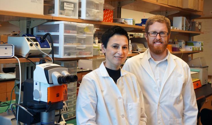
Research scientist Zhanna Nepiyushchikh and principal investigator Brandon Dixon
One of the nasty potential byproducts of surgery to remove cancerous lymph nodes doesn’t rear its ugly head until it’s usually too late to fix.
“During the procedure, some of the lymphatic vasculature is taken out because the surgeon is almost operating blind. If the lymphatic system suffers from injury during surgery, the damage is often difficult to gauge, presenting as lymphedema maybe two to five years later,” says J. Brandon Dixon, associate professor of bioengineering in the Woodruff School of Mechanical Engineering at the Georgia Institute of Technology, where his research is focused on the molecular aspects of lymphatic function in the body’s dynamic, ever-changing mechanical environment.
Dixon, needing an adequate model to study the effects of surgery on the lymphatic system, luckily ran into John Peroni, professor of large animal surgery at the University of Georgia’s (UGA) Regenerative Bioscience Center, during the annual meeting of the Regenerative Engineering and Medicine (REM) research center.
“We started talking about one of the things the lymphatic research community was lacking – a large animal model that could help us understand lymphatic injury,” says Dixon, who also has an appointment in the Wallace H. Coulter Department of Biomedical Engineering at Georgia Tech and Emory. That conversation between he and Peroni has resulted in their recently-published research paper in the journal Nature Biomedical Engineering, “Lymphatic remodelling in response to lymphatic injury in the hind limbs of sheep.”
Dixon and Peroni, who are both members of the Petit Institute for Bioengineering and Bioscience at Georgia Tech, led a multi-institutional collaboration that also included the Albert Einstein College of Medicine (Bronx, N.Y.), and was supported by an REM Seed Grant. Dixon had previously developed imaging technology to capture in vivo lymphatic activity in small animal models, and was eager to test it on a larger animal model that could more accurately reflect the role gravity plays in opposing lymphatic flow in a human.
The main function of the lymphatic system in any vertebrate is to move lymph fluid (loaded with infection-fighting white blood cells) through the body, via lymphatic vessels. There is no central pump, no heart; the lymphatic vessels themselves slowly pump the fluid, and the greatest force working against them is gravity.
One of Dixon’s graduate researchers drove equipment from the Lab of Lymphatic Biology and Bioengineering at Tech to Athens, so Peroni could perform surgery with the near infrared (NIR) image guidance system. The surgery team wanted to compromise the lymphatic vasculature of a sheep in one limb, “so they removed a chain of pumps in one leg and left the other leg intact,” Dixon explains. “Then we followed up with NIR lymphatic imaging on the animal for up to six weeks after the surgical injury. There was an initial decline of pump function, but eventually it started to recover.”
The researchers measured ‘pressure generation capacity’ in the vessels with imaging over the course of remodeling, and then carefully dissected out these sub-millimeter in diameter vessels at the end of six weeks for functional testing. They gauged mechanical properties, performed a proteomic analysis of the muscle cells, and found that the remodeled lymphatic vessel was working much harder than vessels from the control limb.
“Think of it like an elevated heart rate for a lymphatic vessel,” says Dixon, whose team noted, “significantly increased signs of oxidative stress.”
The team found, over the six-week period, that lymphedema hadn’t developed, and it seemed as if the affected lymphatic vessel could handle the load, “but it had to do the work of two vessels, and at the cost of oxidative stress, and that’s analogous to early signs of cardiovascular failure,” notes Dixon.
The stress on the vessel wasn’t the result of the initial surgical injury, “but a result of the vessel’s adaptation for the surgery,” Dixon says. “And it’s a good model for what happens to a human.”
Lead author of the paper was Tyler S. Nelson, former graduate researcher in Dixon lab. Other authors were Zhanna Nepiyushchikh (research scientist in the Dixon lab), Joshua S. T. Hooks and and Mohammad S. Razavi (former graduate researchers in Dixon lab), Tristan Lewis and Merrilee Thoresen (University of Georgia), Cristina Clement and Laura Santambrogio (Einstein College of Medicine), Matthew T. Cribb (graduate researcher in Dixon lab), Mindy K. Ross (former undergraduate researcher in Dixon lab), Rudolph L. Gleason (associate professor in Woodruff School and Coulter Department), Peroni, and Dixon.
Jerry Grillo
Communications Officer II
Parker H. Petit Institute for
Bioengineering and Bioscience


